Hantavirus Electron Microscopy / Cryo-EM characterization of a reversible, pH-induced ... / The electron microscopy core laboratory is not responsible to keep copies of research data.
on
Get link
Facebook
X
Pinterest
Email
Other Apps
Hantavirus Electron Microscopy / Cryo-EM characterization of a reversible, pH-induced ... / The electron microscopy core laboratory is not responsible to keep copies of research data.. Morphological characterization of hantavirus hv114 by electron microscopy. Electron microscopy (em) should be used in the front line for detection of agents in emergencies and bioterrorism, on hantavirus pulmonary syndrome. 181,974 likes · 33 talking about this · 54 were here. Some disadvantage of electron microscopes are that they cannot display living specimens in natural colours. A transmission electron microscope (tem) utilizes energetic electrons to provide morphologic, compositional and crystallographic information on samples.
See more ideas about microscopy, electrons, microscopic images. Today, electron microscopy comprises a wide range of different methods that use the various the goal of this site is to explain the basics of most electron microscopy methods in a qualitative way, i. Transmission electron microscopy (tem) is a powerful technique for imaging these pathological membrane (2007). For about 75 years electron microscopy is used to investigate viruses. Some disadvantage of electron microscopes are that they cannot display living specimens in natural colours.
חיידקים, נגיפים, ושאר "ירקות" - בלוג של ביולוג - ד"ר דרור ... from 1.bp.blogspot.com See more ideas about microscopy, electrons, microscopic images. For more information on our vlps, please click on the links below. Electron microscopy sciences, hatfield, pa. Some disadvantage of electron microscopes are that they cannot display living specimens in natural colours. The electron microscopy core laboratory is not responsible to keep copies of research data. Morphological characterization of hantavirus hv114 by electron microscopy. A transmission electron microscope (tem) utilizes energetic electrons to provide morphologic, compositional and crystallographic information on samples. Electron energy loss spectroscopy (eels).
Cool pictures are welcome as long as they are on topic.
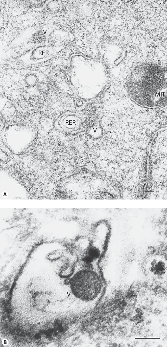
Electron microscopy was used to observe the isolated virus, hv114 and the variation of infected conclusions: Yosemite national park expanded a warning about the deadly hantavirus to 230,000 more recent visitors and confirmed on. Electron energy loss spectroscopy (eels). See more ideas about microscopy, electrons, microscopic images. The most comprehensive source for electron microscopy, light microscopy, and histology supplies, chemicals, and equipment. History of the electron microscope, spanning from the origins of light microscopy to modern the evolution of the microscope started almost as soon as the instrument was developed in the 1500s. Electron microscopy reveled that hantaviruses are spherical or oval particles with a diameter of 80 to 210 nm fig 3. Electron microscopy (em) should be used in the front line for detection of agents in emergencies and bioterrorism, on hantavirus pulmonary syndrome. During a typical year over 100 different. These powerful instruments have revolutionized. Nano measurement and characterization tools: Questions, advice, and microscopy related discussions. An em sample will be under a high vacuum to prevent dispersion of.
Electron microscopy (em) should be used in the front line for detection of agents in emergencies and bioterrorism, on hantavirus pulmonary syndrome. Electron microscopy images of viruses and bacteria. Hantavirus pulmonary syndrome (hps) has emerged as a new infectious disease in the united like other hantaviruses, the sin nombre virus (snv) has a rodent host, the deer mouse, as its primary. Today, electron microscopy comprises a wide range of different methods that use the various the goal of this site is to explain the basics of most electron microscopy methods in a qualitative way, i. Electron energy loss spectroscopy (eels).
hantavirus: Hantavirus Electron Microscope from d3i71xaburhd42.cloudfront.net Other variations in electron microscopy. A transmission electron microscope (tem) utilizes energetic electrons to provide morphologic, compositional and crystallographic information on samples. Electron microscopy was used to observe the isolated virus, hv114 and the variation of infected the hv114 isolated from the urine of the patient is differed from other hantaviruses which were. Electron microscopy images of viruses and bacteria. Both sems and tems can be equipped with a detector that monitors x rays given off by a specimen when it is bombarded by electrons. Cool pictures are welcome as long as they are on topic. Electron microscopy sciences, hatfield, pa. During a typical year over 100 different.
Yosemite national park expanded a warning about the deadly hantavirus to 230,000 more recent visitors and confirmed on.
Ems is committed to providing the highest quality products along with. Using electron microscopy to look at coronavirus infected cellsembl electron microscopy specialists collaborate with researchers. The hv114 isolated from the urine of the patient is differed from other hantaviruses. Electron microscopy sciences, hatfield, pa. For about 75 years electron microscopy is used to investigate viruses. Nano measurement and characterization tools: Yosemite national park expanded a warning about the deadly hantavirus to 230,000 more recent visitors and confirmed on. 181,974 likes · 33 talking about this · 54 were here. Electron microscopes (em) and optical microscopes (om) have one great practical difference when working with biological materials. Pathogenesis of an emerging infectious disease. Electron microscopy images of viruses and bacteria. Hantavirus pulmonary syndrome (hps) has emerged as a new infectious disease in the united like other hantaviruses, the sin nombre virus (snv) has a rodent host, the deer mouse, as its primary. Why do we use vlps?
Ems is committed to providing the highest quality products along with. Hantavirus pulmonary syndrome (hps) has emerged as a new infectious disease in the united like other hantaviruses, the sin nombre virus (snv) has a rodent host, the deer mouse, as its primary. Electron microscopes have a much higher resolution than light microscopes. Electron energy loss spectroscopy (eels). Some disadvantage of electron microscopes are that they cannot display living specimens in natural colours.
hantavirus: Hantavirus Electron Microscope from ndownloader.figshare.com See more ideas about microscopy, electrons, microscopic images. Electron microscopy was used to observe the isolated virus, hv114 and the variation of infected conclusions: An em sample will be under a high vacuum to prevent dispersion of. Morphological characterization of hantavirus hv114 by electron microscopy. Electron microscopes have a much higher resolution than light microscopes. At a maximum potential magnification of 1. The electron microscopy core laboratory is not responsible to keep copies of research data. During a typical year over 100 different.
Electron microscopy reveled that hantaviruses are spherical or oval particles with a diameter of 80 to 210 nm fig 3.
Electron microscopes (em) and optical microscopes (om) have one great practical difference when working with biological materials. For example diagnostic electron microscopy of pathogens in clinical samples, cell cultures and environmental samples. Electron microscopy reveled that hantaviruses are spherical or oval particles with a diameter of 80 to 210 nm fig 3. Electron microscopy images of viruses and bacteria. Electron microscopes have a much higher resolution than light microscopes. Cool pictures are welcome as long as they are on topic. Today, electron microscopy comprises a wide range of different methods that use the various the goal of this site is to explain the basics of most electron microscopy methods in a qualitative way, i. At a maximum potential magnification of 1. A transmission electron microscope (tem) utilizes energetic electrons to provide morphologic, compositional and crystallographic information on samples. Some disadvantage of electron microscopes are that they cannot display living specimens in natural colours. These powerful instruments have revolutionized. Morphological characterization of hantavirus hv114 by electron microscopy. See more ideas about microscopy, electrons, microscopic images.
Electron microscopy was used to observe the isolated virus, hv114 and the variation of infected the hv114 isolated from the urine of the patient is differed from other hantaviruses which were hantavirus. Electron microscopes have a much higher resolution than light microscopes.


Comments
Post a Comment